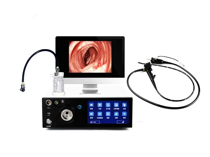
The desktop host of gastrointestinal endoscope is the core control unit of the digestive endoscope system. It is responsible for image processing, light source control, data storage and auxiliary diagnosis. It is widely used in gastroscopy, colonoscopy and other examinations and treatments (such as polypectomy, ESD/EMR surgery). The following are its main components and functional features:
1. Core functional modules
(1) Image processing system
High-definition imaging: supports 1080p/4K resolution, with CMOS or CCD sensors to ensure that mucosal texture and capillaries are clearly visible.
Real-time image optimization:
HDR (high dynamic range): balances bright and dark areas to avoid reflection or loss of dark area details.
Electronic staining (such as NBI/FICE): enhances lesion contrast through narrow-band spectrum (early cancer identification).
AI assistance: automatically marks suspicious lesions (such as polyps, ulcers), and some systems support real-time pathological grading (such as Sano classification).
(2) Light source system
LED/Laser cold light source: adjustable brightness (e.g. ≥100,000 Lux), color temperature adapted to different inspection requirements (e.g. white light/blue light switching).
Intelligent dimming: automatically adjusts brightness according to lens distance to avoid overexposure or insufficient light.
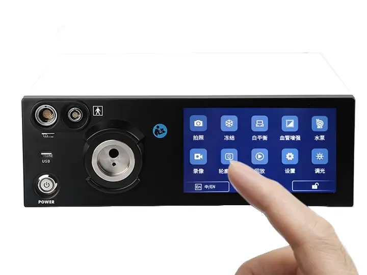
(3) Data management and output
Recording and storage: supports 4K video recording and screenshots, compatible with DICOM 3.0 standard, and can be connected to hospital PACS system.
Remote collaboration: enables real-time consultation or teaching live broadcast through 5G/network.
(4) Treatment function integration
Electrosurgical interface: connects to high-frequency electrosurgical unit (e.g. ERBE) and argon gas knife, supports polypectomy, hemostasis and other operations.
Water injection/gas injection control: integrated regulation of intracavitary water injection and suction to simplify the operation process.
2. Typical technical parameters
Item Parameter example
Resolution 3840×2160 (4K)
Frame rate ≥30fps (smooth without delay)
Light source type 300W Xenon or LED/Laser
Image enhancement technology NBI, AFI (autofluorescence), AI tagging
Data interface HDMI/USB 3.0/DICOM
Sterilization compatibility The host does not require disinfection, and the mirror supports immersion/high temperature
3. Application scenarios
Diagnosis: gastric cancer/intestinal cancer screening, inflammatory bowel disease evaluation.
Treatment: polypectomy, ESD (endoscopic submucosal dissection), hemostatic clip placement.
Teaching: surgical video playback, remote teaching.
Summary
The desktop host of the gastrointestinal endoscope has become the "brain" of digestive endoscopy diagnosis and treatment through high-definition imaging, intelligent image processing and multi-device collaboration. Its technical core lies in image quality, functional scalability and ease of operation. In the future, it will further integrate AI and multimodal imaging technology to improve the early cancer detection rate and surgical efficiency.