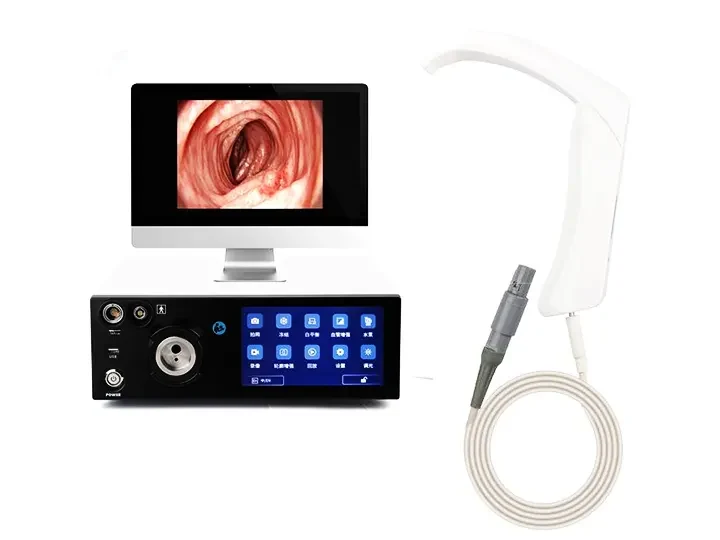
The medical endoscope host is a highly integrated system, mainly composed of an image processing module, a light source system, a control unit and auxiliary accessories to ensure clear endoscope imaging and stable operation.
1. Image processing system
(1) Image processor (video processing center)
Function: Receive endoscope sensor (CMOS/CCD) signals and perform noise reduction, sharpening, HDR enhancement, and color correction.
Technology: Support 4K/8K resolution, low-latency encoding (such as H.265), and AI real-time analysis (such as lesion marking).
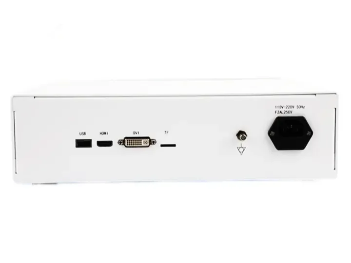
(2) Video output module
Interface type: HDMI, SDI, DVI, etc., connected to a display or recording device.
Split screen function: Supports multi-screen display (such as white light + fluorescence synchronous contrast).
2. Light source system
(1) Cold light source generator
Light source type:
LED light source: energy-saving, long life (about 30,000 hours), adjustable brightness.
Xenon light source: high brightness (>100,000 Lux), color temperature close to natural light.
Intelligent control: Automatically adjust the brightness according to the surgical field (such as brightening the bleeding scene).
(2) Fiber optic interface
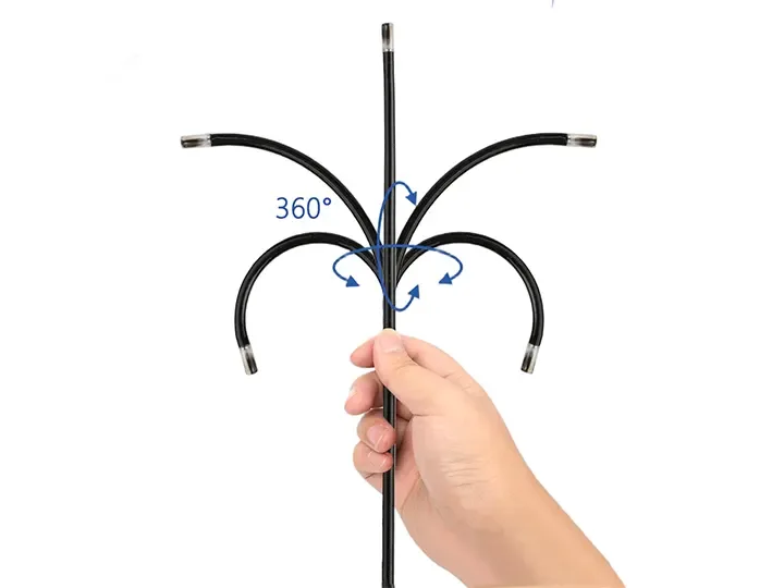
Light guide connector: transmits the light source to the front end of the endoscope to illuminate the inspection area.
3. Control and interaction unit
(1) Main control panel/touch screen
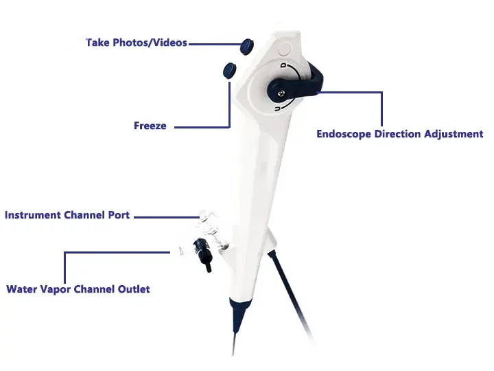
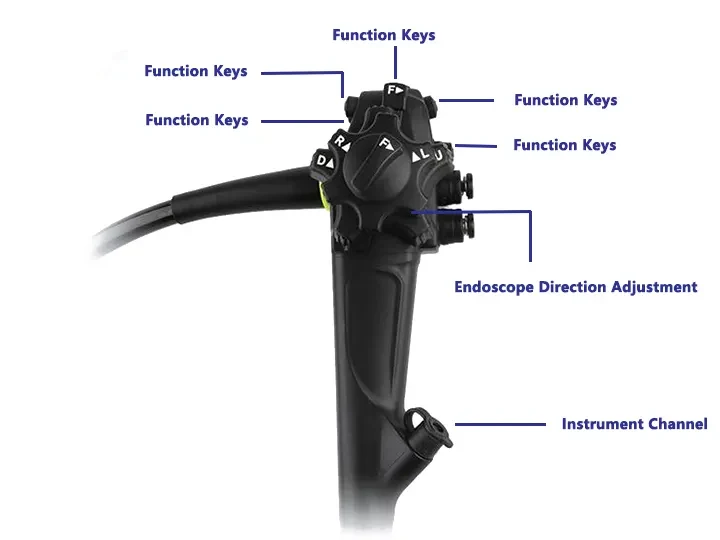
Function: adjust parameters (brightness, contrast), switch imaging mode (NBI/fluorescence), video control.
Design: physical buttons or touch screen, some support voice commands.
(2) Foot switch (optional)
Purpose: Doctors can operate hands-free during surgery, such as freezing images and switching light source modes.
4. Data storage and management module
(1) Built-in storage
Hard disk/SSD: record 4K surgical videos (usually supports more than 1TB capacity).
Cloud synchronization: some hosts support uploading cases to the cloud.
(2) Data interface
USB/Type-C: export case data.
Network interface: remote consultation or access to the hospital PACS system.
5. Auxiliary expansion accessories
(1) Insufflator interface (for laparoscopy only)
Function: connect to insufflator to automatically adjust intra-abdominal air pressure.
(2) Energy device interface
Compatible with high-frequency electrosurgical knife and ultrasonic scalpel: realize electrocoagulation, cutting and other operations.
(3) 3D/fluorescence module (high-end model)
3D imaging: output stereoscopic images through dual cameras.
Fluorescence imaging: such as ICG fluorescence marking tumor boundaries.
6. Power supply and cooling system
Redundant power supply design: prevent power failure during surgery.
Fan/liquid cooling: ensure long-term working stability.